Birthmark Removal: Which Marks Fade With Laser, Which Need Surgery, And Which Should Be Left Alone
<style type="text/css"> </style> Birthmark removal depends on the type. Nevus of Ota and some pigmented marks lighten with a series of Q-switched or picosecond laser sessions, port-wine stains respond to vascular lasers, and raised or large congenital moles may need staged surgical excision. Cafe au lait spots respond unpredictably, and many infant haemangiomas fade on their own or with medicine.
Birthmarks are coloured or raised areas of skin that are present at birth or develop shortly afterwards. Some remain unchanged throughout life, while others grow, darken, fade or change texture.
Parents and adults often ask whether a birthmark can be removed permanently without leaving a scar.
The answer depends on the type of birthmark.
A flat brown patch behaves differently from a blue-grey mark, a red vascular stain or a raised congenital mole.
Choosing treatment before identifying the lesion can result in unnecessary procedures, pigmentation changes or delayed diagnosis of a condition that requires medical attention.
Which Type of Birthmark Do You Have?
Birthmarks are broadly classified as pigmented or vascular.
1. Pigmented Birthmarks
Pigmented birthmarks develop because of changes in the number, location or activity of pigment-producing cells.
Common examples include:
-
Café-au-lait spots
-
Nevus of Ota
-
Congenital melanocytic nevi
-
Dermal melanocytosis
These marks may appear light brown, dark brown, black or blue-grey.
Some are suitable for pigment-targeting lasers, while others require monitoring or surgical assessment.
2. Vascular Birthmarks
Vascular birthmarks involve blood vessels within the skin.
Examples include:
-
Port-wine stains
-
Infantile haemangiomas
-
Certain congenital vascular malformations
These lesions may appear pink, red, purple or bluish.
Their treatment differs from that used for pigmented birthmarks.
Why Diagnosis Matters
A brown birthmark is not automatically a mole, and a red birthmark is not necessarily a haemangioma.
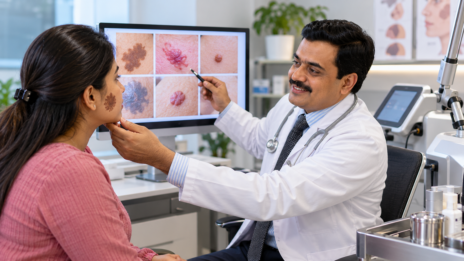
The clinician examines the lesion's colour, depth, texture, location and history.
Dermatoscopy, imaging or other investigations may be required in selected cases.
Changing, bleeding, painful or otherwise suspicious pigmented lesions should be assessed before cosmetic treatment.
Which Birthmarks Respond to Laser, and How Many Sessions?
Different lasers target different structures within the skin.
The appropriate laser depends on whether the birthmark contains excess pigment or abnormal blood vessels.
Birthmark removal laser treatment may use Q-switched or picosecond technology for selected pigmented lesions, while vascular birthmarks often require pulsed dye or other blood-vessel-targeting lasers.
Nevus of Ota: Q-Switched or Picosecond Laser
Nevus of Ota is a blue-grey or brownish pigmentation condition commonly affecting skin around the eye, forehead or cheek.
It occurs when pigment-producing cells are located deeper within the skin.
Nevus of Ota treatment may involve Q-switched or picosecond pigment-targeting lasers.
These lasers deliver short pulses of energy that target pigment.
The body's natural processes gradually clear some of the fragmented pigment.
Multiple sessions are usually required.
Some patients experience substantial lightening, but complete clearance cannot be guaranteed.
Because nevus of Ota can involve the eye, patients should receive an appropriate ophthalmological assessment, including evaluation for ocular pigmentation and glaucoma risk.
Port-Wine Stain: Vascular Laser
A port-wine stain is a capillary malformation that typically appears as a flat pink, red or purple patch.
Unlike many infantile haemangiomas, port-wine stains generally do not disappear spontaneously.
A port wine stain laser treatment commonly uses a pulsed dye laser.
The laser targets haemoglobin within abnormal superficial blood vessels.
Repeated treatments may gradually lighten the mark.
Some lesions respond better than others, and complete clearance is uncommon.
Large facial port-wine stains, particularly those involving the forehead or upper eyelid, may require evaluation for associated conditions such as Sturge-Weber syndrome and glaucoma.
Café-au-Lait Spots: Variable Laser Response
Café-au-lait spots are flat, light-brown patches.
They are usually harmless when isolated.
Some pigment lasers may lighten these marks, but results are unpredictable.
The pigmentation can return, and treatment may occasionally cause darkening or lightening of the surrounding skin.
Multiple café-au-lait spots, especially in children, may warrant medical evaluation for associated genetic conditions.
Laser treatment should not replace that assessment.
Congenital Melanocytic Nevi: Laser Is Not Always Appropriate
Congenital melanocytic nevi are pigmented moles present at birth.
They may be flat, raised, hairy or irregular in appearance.
Laser treatment is generally not the preferred method for suspicious melanocytic lesions because it may leave deeper nevus cells behind and complicate subsequent monitoring.
Management depends on the lesion's size, appearance, location and associated risks.
Observation or surgical excision may be more appropriate.
Birthmark Type-to-Treatment Comparison
| Birthmark type | Colour and texture | Common management | Typical treatment course | Main risks | Observation appropriate? |
|---|---|---|---|---|---|
| Nevus of Ota | Blue-grey or brown, usually flat | Q-switched or picosecond laser | Multiple sessions, often several or more | Pigment changes, incomplete clearance | Sometimes, with appropriate eye assessment |
| Port-wine stain | Flat pink, red or purple patch | Pulsed dye or another vascular laser | Multiple sessions | Bruising, pigment changes, incomplete response | Depends on location and associated risks |
| Café-au-lait spot | Flat light-brown patch | Observation or selected pigment laser | Variable; repeat treatment may be needed | Recurrence, darkening or lightening | Often |
| Congenital melanocytic nevus | Brown or black, flat or raised | Monitoring or selected excision | Depends on lesion size | Surgical scar, recurrence, lesion-specific risks | Often, with specialist guidance |
| Infantile haemangioma | Red or bluish vascular growth | Observation or medication for higher-risk lesions | Individualised | Ulceration, functional impairment, residual skin changes | Often for small, low-risk lesions |
| Dermal melanocytosis | Flat blue-grey patch, often present in infancy | Usually observation | Often fades during childhood | Generally low risk when typical | Usually |
Important: Session counts cannot be predicted from colour alone. Lesion depth, size, skin type, laser settings and response all influence treatment.
When Is Surgical Excision the Better Choice?
Birthmark removal surgery may be considered when the lesion is raised, structurally unsuitable for laser treatment, suspicious or causing functional problems.
Surgery is not automatically required for every congenital mole.
Raised Congenital Moles
Some congenital melanocytic nevi are raised or contain deeper pigment cells.
Excision may be considered for selected lesions based on clinical findings, cosmetic concerns, symptoms or surveillance difficulties.
The decision should include a discussion of scarring and the limitations of surgery.
Large Congenital Birthmarks
Large or giant congenital melanocytic nevi require specialist evaluation.
Their management depends on size, location, appearance and associated health risks.
Some patients undergo staged surgical excision, while others are monitored.
Surgery does not necessarily eliminate all melanoma risk, particularly when nevus cells extend deeply or occur outside the visible skin lesion.
Birthmarks That Affect Function
A lesion may require treatment if it interferes with:
-
Vision
-
Breathing
-
Feeding
-
Normal movement
-
Skin integrity
Functional problems can make treatment more urgent than cosmetic appearance.
Does Surgery Remove a Birthmark Completely?
Surgery can remove some lesions completely, but it creates a wound that heals with a scar.
Larger lesions may require staged procedures, tissue expansion or reconstruction.
Patients should understand that replacing a visible birthmark with a surgical scar may or may not provide a preferable cosmetic result.
Information about related procedures is available in the guide to scar and mole removal surgery.
Why Are Birthmark Removal Creams Risky?
Online products sometimes claim to remove birthmarks without lasers or surgery.
These claims should be approached cautiously.
Birthmarks Are Not All Surface Pigmentation
Many birthmarks involve pigment cells or blood vessels located deeper within the skin.
A cream applied to the surface cannot reliably eliminate these structures.
Possible Birthmark Removal Cream Side Effects
Unregulated or inappropriate products may cause:
-
Skin irritation
-
Chemical burns
-
Allergic reactions
-
Darkening of the skin
-
Unwanted light patches
-
Permanent scarring
-
Delayed diagnosis of suspicious lesions
Some skin-lightening products may also contain harmful or undeclared ingredients.
Why Strong Acids Should Be Avoided
Applying concentrated acids or corrosive substances to a birthmark can injure healthy skin without treating the underlying lesion appropriately.
This is particularly dangerous around the eyes and on children's skin.
Do not attempt to burn, scrape or chemically remove a birthmark at home.
Can Ordinary Pigmentation Creams Help?
Certain prescription creams may be useful for specific acquired pigmentation disorders.
However, these conditions are not the same as congenital birthmarks.
For example, treatment of white patches addresses a different group of skin conditions.
A correct diagnosis is necessary before selecting medication.
Which Birthmarks Should Be Watched Rather Than Removed?
Not every birthmark requires active treatment.
Some fade naturally, while others remain harmless and stable.
Small, Low-Risk Infantile Haemangiomas
Infantile haemangiomas commonly grow during early infancy before gradually involuting over several years.
Small lesions in low-risk locations may only need monitoring.
However, haemangiomas near the eye, nose, lips, airway or other high-risk areas may require early treatment.
Ulcerated, rapidly growing or function-threatening lesions also need prompt assessment.
When treatment is indicated, oral propranolol is commonly used under appropriate medical supervision.
Typical Dermal Melanocytosis
Dermal melanocytosis commonly appears as blue-grey pigmentation in infancy.
Typical lesions often fade during childhood and do not require removal.
Unusual, extensive or persistent lesions may need further assessment.
Stable Café-au-Lait Spots
An isolated café-au-lait spot often needs no treatment.
However, multiple spots or associated symptoms may warrant medical evaluation.
Selected Congenital Moles
Some congenital melanocytic nevi can be monitored rather than removed.
The clinician considers size, appearance, location, changes over time and individual risk factors.
Parents should receive guidance about what changes require reassessment.
Watch-Only Checklist
Observation may be reasonable when:
-
The birthmark has a confirmed benign diagnosis.
-
It is not interfering with normal function.
-
It is not ulcerating or bleeding.
-
It is not changing in a concerning way.
-
There are no associated medical warning signs.
-
A specialist has recommended monitoring.
Observation does not mean ignoring a birthmark. Some lesions require scheduled follow-up or evaluation for associated conditions.
Can Laser Treatment Cause Pigmentation Changes in Indian Skin?
Skin containing more melanin may have a greater risk of pigmentary changes after laser treatment.
These changes include post-inflammatory hyperpigmentation, which causes darkening, and hypopigmentation, which causes light patches.
Why Does Pigmentation Change?
Laser energy is absorbed by target structures within the skin.
If surrounding pigment cells are affected, the treated area may become darker or lighter.
The risk depends on:
-
Skin type
-
Laser wavelength
-
Energy settings
-
Birthmark depth
-
Sun exposure
-
Previous pigmentation problems
-
Individual healing response
How Can Risk Be Reduced?
The clinician may consider conservative settings, appropriate cooling, careful treatment intervals and test spots where suitable.
Sun protection is important before and after treatment.
Patients should also understand that a test spot cannot eliminate all risk.
General information about related procedures is available under laser treatments, although birthmark-specific lasers and resurfacing lasers are not interchangeable.
What Happens During Birthmark Laser Treatment?
Treatment begins with a clinical examination.
The clinician identifies the birthmark type and determines whether laser treatment is appropriate.
Before Treatment
The assessment may include:
-
Reviewing when the mark first appeared.
-
Checking whether its colour, size or texture has changed.
-
Examining its location and depth.
-
Reviewing previous treatments.
-
Assessing skin type and pigmentation risk.
-
Discussing expected results and possible complications.
During Treatment
Protective eyewear is used according to the laser system and treatment location.
The laser delivers controlled energy to the selected area.
Patients may experience a snapping or stinging sensation.
Anaesthesia or other comfort measures may be considered depending on the procedure.
After Treatment
Temporary redness, swelling, darkening or bruising may occur.
Some vascular laser treatments produce purpura, or purple bruising, that can last several days.
Pigment-targeting lasers may cause temporary darkening or crusting.
Patients should follow the clinician's instructions and avoid picking at treated skin.
Multiple sessions may be needed, and results are assessed over time.
Birthmark Surgery Near Bhestan: How Is Treatment Selected?
Patients considering birthmark surgery near bhestan should first have the lesion accurately diagnosed.
At Elegance Clinic, Dr. Ashutosh Shah, MBBS, MS, MCh, DNB, Plastic and Reconstructive Surgeon practising since 2004, can assess the need for surgical removal, reconstructive planning and appropriate specialist referral for birthmark conditions.
The consultation should establish:
-
Whether the mark is pigmented or vascular.
-
Whether it is benign or requires further investigation.
-
Whether treatment is medically necessary.
-
Whether laser, surgery, medication or observation is most appropriate.
-
Whether the lesion is near a sensitive structure.
-
Whether treatment could leave a scar or pigmentation change.
-
How many procedures may be required.
-
Whether a child needs paediatric dermatology or another specialist assessment.
Patients should also discuss whether the expected improvement justifies the risks.
For facial moles, the guide to til hatane ke tareeke provides additional information about removal approaches.
Key Takeaways
-
Birthmark removal laser treatment is suitable for selected birthmarks, not every skin mark.
-
Nevus of Ota may respond to Q-switched or picosecond pigment lasers.
-
Port-wine stains are commonly treated with pulsed dye or other vascular lasers.
-
Café-au-lait spots may lighten with laser but can recur.
-
Congenital melanocytic nevi may require monitoring or surgical assessment.
-
Large or raised birthmarks sometimes need staged excision.
-
Many small, low-risk infantile haemangiomas gradually fade without procedures.
-
High-risk haemangiomas require early specialist evaluation.
-
Birthmark removal creams can cause burns, pigmentation changes and scarring.
-
Laser treatment can cause darkening or lightening of the skin.
-
Complete removal is not always possible, and surgery leaves a scar.
-
Accurate diagnosis is the most important step before treatment.
Conclusion: Laser, Surgery or Observation?
Choosing between birthmark removal laser treatment, surgery and observation depends on the type of lesion rather than its appearance alone.
Some pigmented birthmarks respond to targeted lasers, while vascular lesions require different laser systems.
Selected congenital moles may need surgical removal or ongoing monitoring, and many harmless childhood birthmarks require no active treatment.
Before undergoing any procedure, patients should understand the diagnosis, expected benefits, possible scarring, pigmentation risks and likelihood of recurrence.
The appropriate treatment is the one that addresses the birthmark safely and for a clear medical or cosmetic reason not necessarily the one that removes it most aggressively.